
Fig. 8
- ID
- ZDB-FIG-140321-16
- Publication
- Miller et al., 2000 - sucker encodes a zebrafish Endothelin-1 required for ventral pharyngeal arch development
- Other Figures
- All Figure Page
- Back to All Figure Page
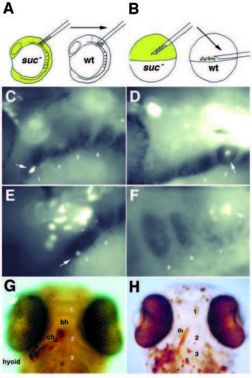
suc/et-1 functions nonautonomously in neural crest and mesoderm. (A) Schematic of neural crest transplants. Premigratory neural crest cells from biotin-labeled donors were tranplanted into unlabeled hosts at the 5 somite stage. (B) Schematic of mesodermal transplants. Cells from the head muscle domain of biotin-labeled gastrulas (Kimmel et al., 1990) were transplanted into the marginal region of unlabeled hosts. (C-H) Genetic mosaic animals at 24 hours (C-F) and 4 days (G,H), with the arches numbered. (C-F) Superimposed fluorescent and bright-field images to colocalize the biotin lineage tracer with dHAND expression in whole-mount embryos. (C,E) suc/et-1 mutant neural crest cells migrate into the dHAND-expressing region of arch one (C) and two (E). In both of these mosaic animals, suc/et-1 mutant ventral neural crest expressed dHAND (see Table 3). Both wild-type (not shown) and suc/et-1 mutant mesoderm (D) fills the non-dHAND-expressing central core in a wild-type host. Initially, neural crest cells lie lateral to mesoderm (F). (G) Mosaic larva in which suc/et-1 mutant neural crest cells, labeled in brown, contributed to a normal ventral cartilage (the ceratohyal) in an unlabeled wild-type host. (H) Mosaic larva in which labeled suc/et-1 mutant mesodermal cells have contributed to a normal ventral muscle (interhyoideus) in a wild-type host. bh, basihyal; ch, ceratohyal; ih, interhyoideus. |