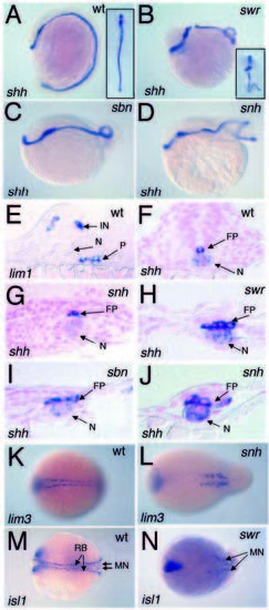
Examination of floor plate and motorneurons in dorsalized mutant embryos. shh expression in wild-type (A), swr/bmp2b (B), sbn/smad5 (C) and snh/bmp7 (D) mutant embryos at the 14-somite stage. The axis of the wild-type embryo (inset A) is straight, while the axis of the swr/bmp2b mutant (inset B) is curved. An intermediate dorsoventral spinal cord position of lim1+ cells in a cross-section of a 14-somite-stage wild-type embryo (E). Crosssections of 14-somite-stage embryos showing floor plate shh expression 1 cell wide in wild-type (F) and 2 to 5 cells wide in snh/bmp7 (G,J), swr/bmp2bsbn/smad5 (I). lim3 expression in the motorneurons of 7-somite-stage wild-type (K) and snh/bmp7 (L) mutant embryo. Expression of isl1 in the RB neurons and motorneurons in wild-type (M) and swr/bmp2b (N), where the motorneurons are apparent, but RB cells are absent. Anterior is to the top, dorsal to the right in A. Anterior is to the left, dorsal to the top in B-D. In K-N, dorsal views, anterior is to the left. FP, floor plate; P, pronephric region.
|
