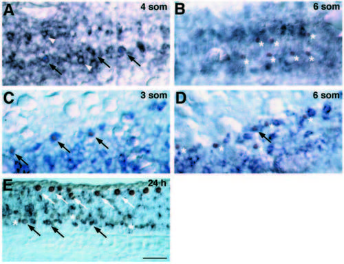
deltaA is transiently expressed in cells specified for neuronal fate. Embryos labeled for nuclear Isl protein (brown), revealing pPMNs and pRBs, and deltaA RNA (blue). (A) Dorsal view of medial neural keel, at the position of the third and fourth somites, at the 4-somite stage (11.3 h). Doubly labeled cells (arrows) indicating pPMNs expressing deltaA. Many cells expressing deltaA at high level do not express Isl (white arrowheads). (B) Similar view to A, at the 6- somite stage. pPMNs (asterisks) do not express deltaA and lie ventral to most cells expressing deltaA at high level. (C) Dorsal view of lateral neural plate, at the position of the 2nd and 3rd somites, of a 3-somite-stage embryo. Doubly labeled pRBs are indicated by arrows. (D) Similar view to C of a 6-somite-stage embryo. Most pPMNS (asterisks) no longer express deltaA RNA. A single doubly labeled cell is indicated by the arrow. (E) Saggital section of 24 h spinal cord. Some presumptive secondary motoneurons express deltaA RNA (black arrows) while others do not (asterisks). RBs (white arrows) do not express deltaA at this stage. Scale bar, 25 μm.
|
