
Fig. 4
- ID
- ZDB-FIG-140305-73
- Publication
- Appel et al., 1998 - Regulation of neuronal specification in the zebrafish spinal cord by Delta function
- Other Figures
- All Figure Page
- Back to All Figure Page
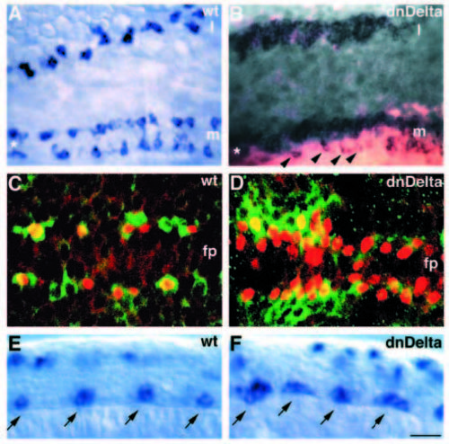
Disruption of Delta function causes overproduction of primary motoneurons. (A) Dorsal view of neural plate of an uninjected 4-somite-stage embryo showing islet1 expression by in situ RNA hybridization in medial (m) and lateral (l) regions. islet1 expression marks pPMNs medially (two longitudinal rows bordering the floorplate, marked by an asterisk) and pRBs laterally. (B) Dorsal view of 4-somite-stage embryo injected with X-Delta-1STU and lacZ mRNA and probed for islet1 expression. Excess pPMNs and pRBs develop medially and laterally, respectively, on the side receiving most injected mRNA. Arrowheads indicate distribution of motoneurons, which is similar to that of uninjected embryos, on the side receiving little mRNA. Asterisk marks position of floorplate between motoneuron rows. (C) Confocal image, dorsal view, of uninjected 6-somite-stage embryo labeled with α-Isl (red) and a-Hu (green) antibodies. α-Isl reactivity precedes that of α-Hu (data not shown); thus, α-Isl+ motoneurons flanking the floorplate (fp) are sometimes unlabeled by α-Hu. Several non-motoneurons (a-Hupositive, α-Isl-negative) are interspersed among motoneurons. (D) Confocal image, dorsal view, of 6-somite-stage embryo injected with X-Delta-1STU mRNA and labeled with α-Isl (red) and α-Hu (green). Nearly every cell flanking the floorplate is a motoneuron. Some motoneurons also develop just dorsal to the floorplate. (E) Wild-type expression pattern of islet2 RNA at 16-somite-stage, shown in lateral view. islet2 is expressed in CaP and VaP motoneurons (arrows). Labeled cells in dorsal spinal cord are pRBs. (F) Similarly staged embryo injected with X-Delta-1STU mRNA and probed for islet2 expression. islet2 is expressed in clusters of cells located at the normal positions of CaP and VaP motoneurons (arrows). Scale bar, 20 μm (A,B), 10 μm (C,D) and 15 μm (E,F). |