
Fig. 2
- ID
- ZDB-FIG-140203-2
- Publication
- Novorol et al., 2013 - Microcephaly models in the developing zebrafish retinal neuroepithelium point to an underlying defect in metaphase progression
- Other Figures
- All Figure Page
- Back to All Figure Page
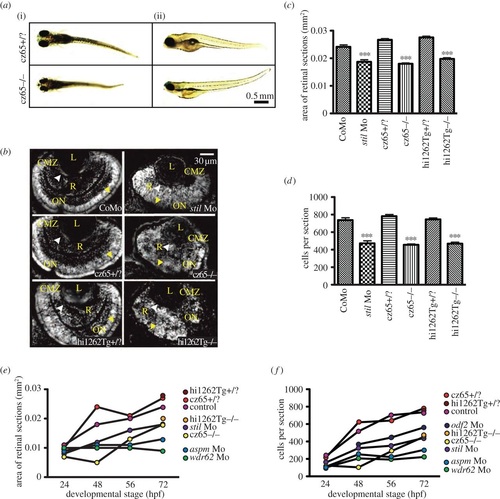
MCPH phenotypes in the zebrafish retina. Knockdown of MCPH genes causes a reduction in head size, retinal size and retinal cell number. (a) Reduction in head and eye size is demonstrated in whole-mount stilcz65-/- mutant embryos on day 5 of development. Healthy stilcz65+/? embryos are shown at the same developmental stage for comparison. While head and eye size were consistently reduced, overall body size/length of mutant embryos was variably affected. Here, one mutant (i) is smaller than the healthy embryo, whereas the other (ii) is similar in length and size to the healthy embryos. Note also the abnormally protruding lenses in the mutant embryo seen from (i), exposed owing to reduced retinal size. (b) DAPI-stained sections demonstrate reduced retinal area in stil morphants (stil Mo) at 72 hpf when compared with control morphants (CoMo). Similarly, the retinas of stilcz65-/- and stilhi1262Tg-/- mutant embryos at 72 hpf are markedly reduced in size. Labels in yellow: L, lens; R, retinal neuroepithelium; CMZ, ciliary marginal zone; ON, optic nerve; AM, apical membrane. (c) Retinal area is significantly reduced in stil morphant and mutant embryos: stil Mo 0.019 mm2 (n = 23) versus CoMo 0.024 mm2 (n = 34), p < 0.001; stilcz65-/- 0.019 mm2 (n = 23) versus stilcz65+/? 0.027 mm2 (n = 72) p < 0.001; stilhi1262Tg-/- 0.020 mm2 (n = 52) versus stilhi1262+/? 0.028 (n = 48), p < 0.001 (values are for mean area at 72 hpf). (d) Retinal cell number is reduced in stil morphants and mutants: stil Mo 471 cells (n = 23) versus CoMo 735 cells (n = 7), p < 0.001; stilcz65-/- 454 cells (n = 114) versus stilcz65+/? 780 cells (n = 72), p < 0.001; stilhi1262Tg-/- 468 cells (n = 52) versus stilhi1262+/? 744 cells (n = 48), p < 0.001 (values are for mean number of cells in central retinal sections at 72 hpf). (e) Retinal area increases as development progresses in stil, aspm, wdr62 and odf2 morphant embryos but remains reduced compared with control at all time-points examined (24, 48, 56 hpf at 72 hpf). (f) Retinal cell increases as development progresses in stil, aspm, wdr62 and odf2 morphant embryos but remains reduced compared with control at all time-points examined (24, 48, 56 hpf at 72 hpf), n = number of eyes analysed. |
| Fish: | |
|---|---|
| Knockdown Reagents: | |
| Observed In: | |
| Stage Range: | Prim-5 to Protruding-mouth |