FIGURE

Fig. S7

ID
ZDB-FIG-131218-37
Publication
Targoff et al., 2013 - Nkx genes are essential for maintenance of ventricular identity
Other Figures
All Figure Page
Back to All Figure Page
Fig. S7

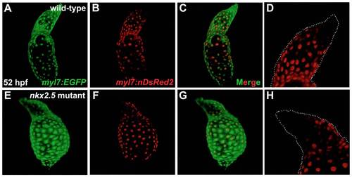
Developmental timing assay indicates late-differentiating cells added to the arterial pole of the nkx2.5 mutant heart.
Confocal projections of hearts in live wild-type (A-D) and nkx2.5 mutant (E-H) embryos expressing Tg(myl7:EGFP) (A,E) and Tg(-5.1myl7:nDsRed2) (B,D,F,H). Lateral views, arterial pole to the top, at 52 hpf. (D,H) White dots outline the morphology of the Tg(myl7:EGFP)-expressing ventricle and outflow tract. (A-D) In the wild-type heart, the late-differentiating cardiomyocyte population exhibits green, but not red, fluorescence, due to the delay in expression of Tg(-5.1myl7:nDsRed2), in comparison with expression of Tg(myl7:EGFP), at the arterial pole (de Pater et al., 2009). (F-H) Similarly, in the nkx2.5 mutant heart, cardiomyocytes expressing Tg(myl7:EGFP), but not Tg(-5.1myl7:nDsRed2), are present at the arterial pole.

Expression Data

Expression Detail
Antibody Labeling
Phenotype Data

Phenotype Detail
Acknowledgments
This image is the copyrighted work of the attributed author or publisher, and ZFIN has permission only to display this image to its users. Additional permissions should be obtained from the applicable author or publisher of the image. Full text @ Development