
|
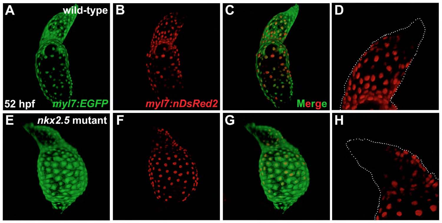
Fig. S7
Developmental timing assay indicates late-differentiating cells added to the arterial pole of the nkx2.5 mutant heart.
Confocal projections of hearts in live wild-type (A-D) and nkx2.5 mutant (E-H) embryos expressing Tg(myl7:EGFP) (A,E) and Tg(-5.1myl7:nDsRed2) (B,D,F,H). Lateral views, arterial pole to the top, at 52 hpf. (D,H) White dots outline the morphology of the Tg(myl7:EGFP)-expressing ventricle and outflow tract. (A-D) In the wild-type heart, the late-differentiating cardiomyocyte population exhibits green, but not red, fluorescence, due to the delay in expression of Tg(-5.1myl7:nDsRed2), in comparison with expression of Tg(myl7:EGFP), at the arterial pole (de Pater et al., 2009). (F-H) Similarly, in the nkx2.5 mutant heart, cardiomyocytes expressing Tg(myl7:EGFP), but not Tg(-5.1myl7:nDsRed2), are present at the arterial pole.