
FIGURE
Fig. 3
- ID
- ZDB-FIG-131213-59
- Publication
- Dohn et al., 2013 - Planar cell polarity proteins differentially regulate extracellular matrix organization and assembly during zebrafish gastrulation
- Other Figures
- All Figure Page
- Back to All Figure Page
Fig. 3
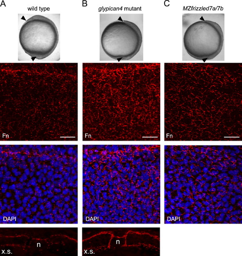
glypican4 and frizzled7 mutant embryos have increased fibronectin assembly. (A–C) Top panels show morphological convergence and extension phenotypes at tailbud stage (see arrowheads) of wild type, glypican4 mutant, and MZfrizzled7a/7b mutant embryos. Middle two panels show confocal images of fibronectin (Fn) immunolabeling without and with nuclear DAPI staining. Scale bars=20 μm. The bottom images in (A) and (B) show Fn expression in cross-sections (x.s.) of tailbud stage embryos (n, notochord). |
Expression Data
Expression Detail
Antibody Labeling
Phenotype Data
| Fish: | |
|---|---|
| Observed In: | |
| Stage: | Bud |
Phenotype Detail
Acknowledgments
This image is the copyrighted work of the attributed author or publisher, and
ZFIN has permission only to display this image to its users.
Additional permissions should be obtained from the applicable author or publisher of the image.
Reprinted from Developmental Biology, 383(1), Dohn, M.R., Mundell, N.A., Sawyer, L.M., Dunlap, J.A., and Jessen, J.R., Planar cell polarity proteins differentially regulate extracellular matrix organization and assembly during zebrafish gastrulation, 39-51, Copyright (2013) with permission from Elsevier. Full text @ Dev. Biol.