
Fig. 7
- ID
- ZDB-FIG-131213-63
- Publication
- Dohn et al., 2013 - Planar cell polarity proteins differentially regulate extracellular matrix organization and assembly during zebrafish gastrulation
- Other Figures
- All Figure Page
- Back to All Figure Page
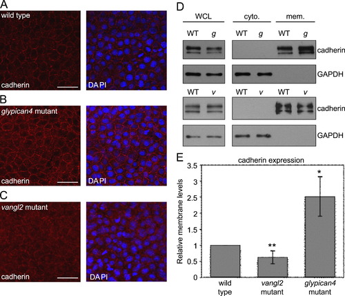
Glypican4 mutant embryos have increased levels of membrane cadherin protein. (A–C) Confocal images of cadherin expression (without or with nuclear DAPI staining) in tailbud stage wild type, glypican4 mutant, and vangl2 mutant embryos. Scale bars=20 μm. (D) Western blot analysis of cadherin expression in embryonic cytoplasmic (cyto.) and membrane (mem.) protein fractions. Cadherin and GAPDH expression in whole cell lysates (WCL) prior to fractionation are also shown. WT, g, and v denote protein extracts obtained from wild type, glypican4 mutant, and vangl2 mutant embryos, respectively. (E) Quantification of western blot fractionation data from triplicate experiments displayed as relative membrane cadherin expression normalized to GAPDH levels in WCL (**p<0.02, *p<0.009). |
Reprinted from Developmental Biology, 383(1), Dohn, M.R., Mundell, N.A., Sawyer, L.M., Dunlap, J.A., and Jessen, J.R., Planar cell polarity proteins differentially regulate extracellular matrix organization and assembly during zebrafish gastrulation, 39-51, Copyright (2013) with permission from Elsevier. Full text @ Dev. Biol.