
FIGURE
Fig. 6
- ID
- ZDB-FIG-131107-25
- Publication
- Lee et al., 2013 - Moving domain computational fluid dynamics to interface with an embryonic model of cardiac morphogenesis
- Other Figures
- All Figure Page
- Back to All Figure Page
Fig. 6
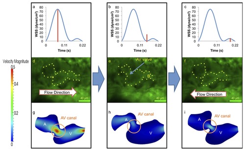
At 110-120 hpf, the morphologic AV valve is distinct and left ventricle is enlarging. (a–c) WSS across AV valve in response to atrium contraction is significantly higher than that of ventricular contraction. (d–i) Complete formation of the AV valve and bulbus arteriosus was observed at this stage, and amount of flow reversal reduced. The ventricle has become larger than the atrium in size, and the flow reversal through the AV canal during ventricular contraction is reduced due to small size of AV canal. A, atrium; V, ventricle; B, bulbus arteriosus. |
Expression Data
Expression Detail
Antibody Labeling
Phenotype Data
Phenotype Detail
Acknowledgments
This image is the copyrighted work of the attributed author or publisher, and
ZFIN has permission only to display this image to its users.
Additional permissions should be obtained from the applicable author or publisher of the image.
Full text @ PLoS One