
Fig. 1
- ID
- ZDB-FIG-131105-10
- Publication
- Lee et al., 2013 - Moving domain computational fluid dynamics to interface with an embryonic model of cardiac morphogenesis
- Other Figures
- All Figure Page
- Back to All Figure Page
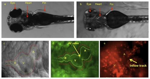
Dorsal view of zebrafish embryos illustrated cardiac system prior to and after pigmentation removal. (a) Pigmentation opacifies internal organ systems at 120 hpf. (b) Treatments with PTU beginning at 10 hpf prevented pigment formation, allowing for clear organ visualization. (c) PTU-treated zebrafish heart observed under bright field microscope at 120 hpf. Despite visualization of outer wall, inner wall is poorly demarcated. (d) The use of transgenic Tg(fli1a:EGFP)y1 embryos allows for clear delineation of inner wall for reconstructing the CFD model. (e) Tg(gata1:dsRed)sd2 transgenic zebrafish allows to visualize the blood particle in order to take velocity by particle tracking technique. A, atrium; V, ventricle; B, bulbus arteriosus. |