
Fig. 4
- ID
- ZDB-FIG-131107-17
- Publication
- Lee et al., 2013 - Moving domain computational fluid dynamics to interface with an embryonic model of cardiac morphogenesis
- Other Figures
- All Figure Page
- Back to All Figure Page
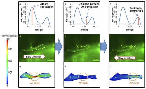
At 20-30 hpf, zebrafish heart is morphologically a tube-like structure with a AV canal (AV) separating the atrium (A) and ventricle (V). (a) Atrial contraction, the first peak, engenders an increase in shear stress profiles through the AV canal. (b) Diastasis between atrial and ventricular contraction result in a decrease in shear stress across the AV canal. (c) Ventricular contraction, the second peak, results in flow reversal or regurgitation through the AV canal. The magnitude of wall shear stress (WSS) across the AV canal is about half of the forward blood flow at AV canal. (d) The green fluorescent protein (GFP) delineated the tubular structure. Unidirectional forward flow from the contracting atrium into the ventricle develops through the AV canal. (e) The GFP image reveals movement of peristaltic wave to the end of tubular heart structure. (f) Flow reversal develops in response to the contraction of tubular structure. (g) Velocity profiles show the maximal magnitude through the AV canal. (h) The magnitude of velocity is minimal at AV canal. (i) The magnitude of flow reversal or regurgitation is about half of the forward blood flow at AV canal. Contoured velocity profiles confirm the observed result (g–i). Red bars in (a–c) represent the time points at which the corresponding velocity profiles were reconstructed. A, atrium; V, ventricle. |