FIGURE

Fig. S3

ID
ZDB-FIG-131106-5
Publication
Hess et al., 2012 - Intravital Imaging of Thymopoiesis Reveals Dynamic Lympho-Epithelial Interactions
Other Figures
All Figure Page
Back to All Figure Page
Fig. S3

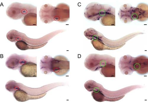
Expression domains of ccl25 and cxcl12 chemokine genes

ÿShown are photographs of 3 dpf wild-type embryos subjected to WISH using gene specific probes. At the top, left images are lateral, right images are dorsal views of embryos. The bottom panels depict the entire embryo (composites of two [A], three [B,C] or four [D] original images).
(A) Expression pattern of ccl25a. The region of the thymus is marked with a red circle.
(B) Expression pattern of ccl25b. The region of the thymus is marked with a red circle.
(C) Expression pattern of cxcl12a. The region of the relevant gill arches encompassing the thymic rudiment is marked with a green circle.
(D) Expression pattern of cxcl12b. The region of the relevant gill arches encompassing the thymic rudiment is marked with a green circle.
Photographs representative of at least 60 embryos per probe. Scale bars, 50μm.

Expression Data
Genes:
Fish:
Anatomical Terms:
Stage: Protruding-mouth

Expression Detail
Antibody Labeling
Phenotype Data

Phenotype Detail
Acknowledgments
This image is the copyrighted work of the attributed author or publisher, and ZFIN has permission only to display this image to its users. Additional permissions should be obtained from the applicable author or publisher of the image.

Reprinted from Immunity, 36(2), Hess, I., and Boehm, T., Intravital Imaging of Thymopoiesis Reveals Dynamic Lympho-Epithelial Interactions, 298-309, Copyright (2012) with permission from Elsevier. Full text @ Immunity