
|
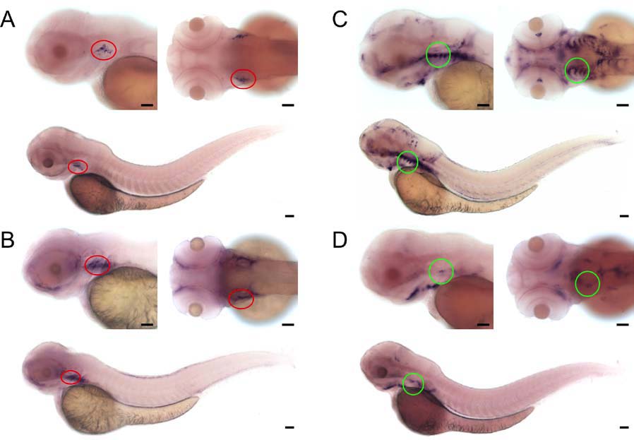
Fig. S3
Expression domains of ccl25 and cxcl12 chemokine genes
ÿShown are photographs of 3 dpf wild-type embryos subjected to WISH using gene specific probes. At the top, left images are lateral, right images are dorsal views of embryos. The bottom panels depict the entire embryo (composites of two [A], three [B,C] or four [D] original images).
(A) Expression pattern of ccl25a. The region of the thymus is marked with a red circle.
(B) Expression pattern of ccl25b. The region of the thymus is marked with a red circle.
(C) Expression pattern of cxcl12a. The region of the relevant gill arches encompassing the thymic rudiment is marked with a green circle.
(D) Expression pattern of cxcl12b. The region of the relevant gill arches encompassing the thymic rudiment is marked with a green circle.
Photographs representative of at least 60 embryos per probe. Scale bars, 50μm.
Reprinted from Immunity, 36(2), Hess, I., and Boehm, T., Intravital Imaging of Thymopoiesis Reveals Dynamic Lympho-Epithelial Interactions, 298-309, Copyright (2012) with permission from Elsevier. Full text @ Immunity