
FIGURE
Fig. S1
Fig. S1
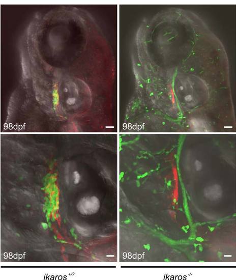
Initial colonization of the thymic rudiment in embryos depends on ikaros function Projected images of fish double-transgenic for ikaros:eGFP and foxn1:mCherry experiments constructs were obtained at 98 hpf. Representative wild-type (ikaros+/?; n=20) and ikaros-deficient (ikaros-/-; n=12) embryos are shown. The green cells seen in the images of the mutant embryos are motile myeloid cells, which do not reside in the thymus and hence do not obscure the view onto the thymic epithelial rudiment (red). For further details see Figure 2 and text. Scale bars, 50μm for upper, and 20μm for lower panels. |
Expression Data
Expression Detail
Antibody Labeling
Phenotype Data
| Fish: | |
|---|---|
| Observed In: | |
| Stage: | Day 4 |
Phenotype Detail
Acknowledgments
This image is the copyrighted work of the attributed author or publisher, and
ZFIN has permission only to display this image to its users.
Additional permissions should be obtained from the applicable author or publisher of the image.
Reprinted from Immunity, 36(2), Hess, I., and Boehm, T., Intravital Imaging of Thymopoiesis Reveals Dynamic Lympho-Epithelial Interactions, 298-309, Copyright (2012) with permission from Elsevier. Full text @ Immunity