
Fig. 1
- ID
- ZDB-FIG-130910-15
- Publication
- Omae et al., 2013 - Identification of Inter-Organ Vascular Network: Vessels Bridging between Organs
- Other Figures
- All Figure Page
- Back to All Figure Page
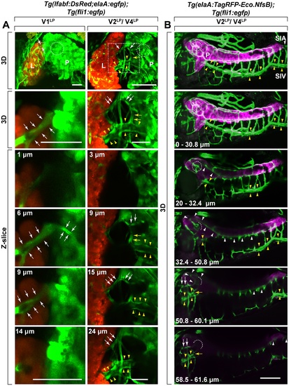
Identification of V1LP, V2LP and V4LP, as inter-organ vessels bridging between liver and pancreas. Three distinct vessels (V1LP, V2LP, V4LP) were found to bridge between liver (L) and pancreas (P). A. The 3D rendered two-photon miscroscopy images (top two panels: 3D) and their serial optical sections (bottom panels: Z-slice) of Tg(lfabf:DsRed;elaA:egfp);Tg(fli1:egfp) at 5 dpf and 6 dpf. The second row 3D panels are the higher magnification of the indicated area (dotted rectangle) in the first row 3D panels. The bottom four panels are Z-slices of the areas shown in second row 3D panels. The depth of each slice is indicated as µm at the top left in each panel of Z-slice. V1LP and V2LP vessels are sandwiched between white arrows and indicated by white arrows, respectively. V4LP vessel is indicated by yellow arrows and supraintestinal veins (SIV) is sandwiched between yellow arrowheads). The islet of Langerhans is indicated (dotted circle in each panel). The islet of Langerhans, consisting of endocrine cells, was identified as an area devoid of EGFP reporter signals driven by the exocrine pancreas-specific elaA promoter (see Fig. 2). In the first column, V1LP vessel (sandwiched white arrows) bridging between liver (L) and pancreas (P) can be clearly seen. Examining the series of Z-slices confirms that V1LP invades into liver tissue. In the second column, V2LP (white arrows) and V4LP (yellow arrows and sandwiched between yellow arrowheads) vessels bridging between liver (L) and pancreas (P) are shown. A connection between V4LP and V2LP appears to exist (yellow arrows in the 2nd panel). Liver (L: orange); endocrine pancreas (P: green); fli1+ vessels (green). Scale bars: 30 μm. B. V2LP (white arrows) and V4LP (yellow arrows and sandwiched between yellow arrowheads) vessels originate from supraintestinal arteries (SIA) and supraintestinal veins (SIV), respectively. The origins of V2LP and V4LP vessels were followed by using Tg(elaA:TagRFP-Eco.NfsB);Tg(fli1:egfp) at 6 dpf. The 3D-rendered Z-stack confocal microscopy images of several Z-slices (depth of ranges is indicated at the bottom left in each panel) are shown in series. Fli1+ vessels and elaA+ exocrine pancreas are shown as green and magenta, respectively. By following the SIA (white arrowheads) in each Z-stack, V2LP is found to originate from vascular plexus at the islet of Langerhans (dotted circle) that is formed by branches of SIA. The 4th and 5th Z-stack panels show that vascular plexus (white arrowheads) at the islet of Langerhans (dotted circle) is formed by branches of SIA. In the 5th Z-slice panel, the direct connection between this SIA-derived vascular plexus at the islet of Langerhans (dotted circle) and V2LP (white arrows) can be seen. Following the vessels pointed by yellow arrows in each Z-stack demonstrate that V4LP vessel is a part of SIV running ventral to pancreas. Supraintestinal artery (SIA) and supraintestinal vein (SIV) are indicated by white arrowheads and sandwiched between yellow arrowheads, respectively. Scale bars: 100 μm. |