
FIGURE
Fig. S1
- ID
- ZDB-FIG-130910-14
- Publication
- Omae et al., 2013 - Identification of Inter-Organ Vascular Network: Vessels Bridging between Organs
- Other Figures
- All Figure Page
- Back to All Figure Page
Fig. S1
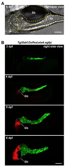
Growth of liver and pancreas between 3 dpf and 6 dpf in developing zebrafish. A. A zebrafish larva at 6 dpf imaged from the right side of the body showing the relative positions of liver (L), pancreas (P), gallbladder (Gb), swimbladder (Sb) and intestine (Int). The anterior and posterior sides on the left and right, respectively. B. The growth of pancreas and liver. The liver (orange) and pancreas (green) were visualized at 3, 4, 5 and 6 dpf using Tg(lfabf:DsRed;elaA:egfp). Gb: Gallbladder. Scale bars: 100 μm. |
Expression Data
Expression Detail
Antibody Labeling
Phenotype Data
Phenotype Detail
Acknowledgments
This image is the copyrighted work of the attributed author or publisher, and
ZFIN has permission only to display this image to its users.
Additional permissions should be obtained from the applicable author or publisher of the image.
Full text @ PLoS One