
Fig. 9
- ID
- ZDB-FIG-130906-31
- Publication
- Sorrell et al., 2013 - Tcf7l1 proteins cell autonomously restrict cardiomyocyte and promote endothelial specification in zebrafish
- Other Figures
- All Figure Page
- Back to All Figure Page
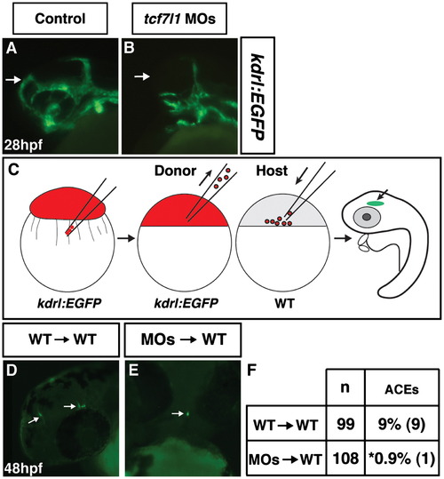
Tcf7l1s cell autonomously repress anterior endothelial cell development. ((A)and (B)) Control and Tcf7l1 deficient Tg(kdrl:EGFP) embryos. Tcf7l1 deficient Tg(kdrl:EGFP) embryos have a dramatic loss of the ACEs at 28 hpf. (C) Schematic of the cell transplantation strategy used for assessing cell autonomy. Red indicates injection with the rhodamine-dextran lineage tracer. (D) Representative image of cranial ECs from donor WT/Tg(kdrl:EGFP) cells transplanted into a WT host. View is fronto-lateral. (E) Representative image of cranial ECs from donor Tcf7l1 deficient/Tg(kdrl:EGFP) cells transplanted into a WT host. View is frontal. (F) Frequency of ACEs found in transplantation experiments. Asterisk indicates a statistically significant difference compared to control transplant experiments. |
| Gene: | |
|---|---|
| Fish: | |
| Knockdown Reagents: | |
| Anatomical Term: | |
| Stage: | Prim-5 |
| Fish: | |
|---|---|
| Knockdown Reagents: | |
| Observed In: | |
| Stage: | Prim-5 |
Reprinted from Developmental Biology, 380(2), Sorrell, M.R., Dohn, T.E., D'Aniello, E., and Waxman, J.S., Tcf7l1 proteins cell autonomously restrict cardiomyocyte and promote endothelial specification in zebrafish, 199-210, Copyright (2013) with permission from Elsevier. Full text @ Dev. Biol.