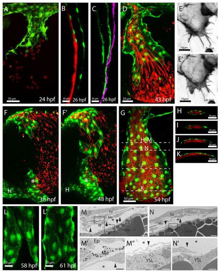
ECs of the CCVs form an open-ended tube before the onset of blood flow and consecutively extend this open vessel. The ECs form an open-ended tube with blood exiting the EC-lined lumen (A,D); the lumen becomes extended by active EC migration (E,E′), during which ECs migrate as a sheet underneath the epidermis (C) and connect to ECs at the heart inflow tract (asterisk, F,F′). Consecutively, the ECs engulf the blood-filled lumen (I,N) and fuse on the YSL side (L,L′). (A-L′) Confocal projections of the developing CCVs, lateral views (A,D,E,G,L), sagittal views (B,C), lateral-ventral view (F) and transverse views (H-K). (D,E,F,L) Projections taken from confocal time-lapse movies. The developing vasculature was visualized by transgenic EGFP expression of Tg(kdrl:EGFP)s843 (B,D,F,G), Tg(etsrp:EGFP)ci1 (A,L), by transgenic mCherry expression of Tg(kdrl:HsHRAS-mCherry)s896 (green in C) or by vascular-specific lifeact-GFP expression [Tg(UAS:lifeact-GFP)mu271 Tg(fli1ep:GAL4FF)ubs4] (E). The YSL was labeled by injection of mini-Ruby into the YSL (B). Erythrocytes are in red, visualized by transgenic dsRed expression of Tg(gata1:dsRed)sd2 (A,D,F-K). The epidermis is in purple [visualized by transgenic EGFP-caax expression of Tg(krt4:EGFP-caax)] (C). Note that the ECs underneath the epidermis extend the CCVs further (C) than on top of the YSL (B). ECs migrate actively with Actin-rich lamellipodia at their front (E,E′). (G-K) Ensheathment of the lumen by ECs; white bars in G indicate the approximate levels of the transverse views shown in H-K,M,N. (L,L′) Lumen closure on the YSL side. ECs of the epidermal side are cropped away. (M-N′) Electron micrograph (higher magnifications, M′,M′,N′) of the CCV of a 48 hpf embryo (scale bars: 10 μm). Arrowheads label ECs, asterisks the CCV lumen. Underneath the epidermis, ECs are next to melanocytes. On the other side of the lumen, ECs are located directly above the YSL. Note that in N there is no complete EC layer above the YSL. Ep, epidermis; Me, melanocytes; YSL, yolk syncytial layer.
|
