
FIGURE
Fig. S4
Fig. S4
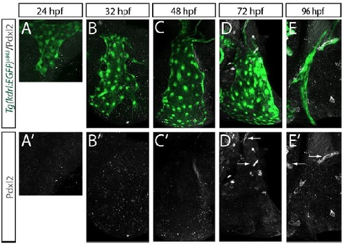
ECs of the CCV have no apical polarization (analyzed by Podocalyxin-like 2 expression). (A-E′) Fluorescent confocal images of immunostainings of Podocalyxin 2 (Pdxl2) at the indicated time points; lateral views. The developing vasculature is visualized by transgenic EGFP expression of Tg(kdrl:EGFP)s843. ECs of the CCV do not form Pdxl2-positive membrane compartments, whereas ECs in the fin bud (arrows in D′ and right arrow in E′) and ECs in the aortic arches (left arrow in E′) form apical membrane compartments as indicated by Pdxl2 staining. |
Expression Data
Expression Detail
Antibody Labeling
Phenotype Data
Phenotype Detail
Acknowledgments
This image is the copyrighted work of the attributed author or publisher, and
ZFIN has permission only to display this image to its users.
Additional permissions should be obtained from the applicable author or publisher of the image.
Full text @ Development