
Fig. S2
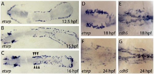
etsrp is strongly expressed in newly specified angioblasts but becomes downregulated after angioblast migration. (A-D,F) Brightfield images showing expression of etsrp (etv2) detected by in situ hybridization in embryos at the indicated time points (dorsal views). (A-C) Flat-mounted embryos; (D-G) higher magnification of the trunk region. (E,G) Brightfield images showing expression of cadherin 5 (cdh5) detected by in situ hybridization in embryos at the indicated time points (dorsal views). (A) etsrp mRNA can be detected in angioblasts within the lateral plate mesoderm. (B) etsrp mRNA levels are lower in angioblasts that have already migrated to the midline. (C) High etsrp expression in angioblasts that will form the bilateral cardinal veins/CCVs (arrowheads). (D) Cells forming the bilateral cardinal veins/CCVs show high levels of etsrp expression, whereas etsrp expression is very weak in the ECs of the LDA. (F) etsrp expression in the bilateral cardinal veins/CCVs decreases over time until it is no longer detectable. (E,G) cdh5 expression labels all angioblasts. CCV, common cardinal vein; LDA, lateral dorsal aortae. |