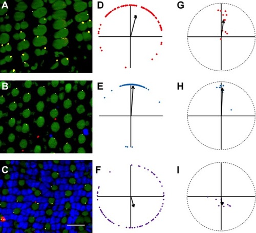
Loss of rods does not affect basal body positioning in cones. Sample portions of fields of cells at the depths of the basal bodies of red-/green-sensitive cones (A), blue-sensitive cones (B), and UV-sensitive cones (C) show basal body positioning in adult light-adapted XOPS-mCFP zebrafish retinas. D-F: Graphs of the positions of basal bodies from fields A-C in which the positions of all the basal bodies of red-/green-, blue-, or UV-sensitive cones were plotted around a unit circle (red, blue, or purple lozenges, respectively), and the mean vector is indicated (arrow). G-I: Mean vectors of basal bodies from individual fields of red-/green-, blue-, and UV-sensitive cones are plotted (red, blue, and purple lozenges, respectively). The grand mean vector of these mean vectors is indicated (arrow). Optic nerve is upward in all panels. Small numbers of remaining rods are occasionally visible (red). β-Tubulin (yellow) localizes to basal bodies. Nuclei were stained with DAPI (blue). Autofluorescence at 488 nm (green) shows inner and outer segments. All fields analyzed were from the middle retina. A magenta-green version of this figure is provided in the Supporting Information. Scale bar = 10 μm.
|
