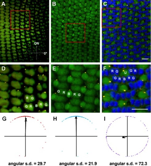
Basal body positioning is strongly patterned in individual fields of red-/green- and blue-sensitive cones. Sample portions of fields of cells at the depths of the basal bodies of red-/green-sensitive cones (A), blue-sensitive cones (B), and UV-sensitive cones (C), along with their magnified subsets of cells (D-F), in adult light-adapted zebrafish retinas. γ-Tubulin localized to basal bodies (yellow), whereas nuclei were counterstained with DAPI (blue). Autofluorescence (green) from 488-nm excitation shows inner and outer segments. Some red-, green-, blue-, and UV-sensitive cones are labeled as R, G, B, and U, respectively. G-I: Graphs of the positions of basal bodies from fields A–C in which the positions of all the basal bodies of red-/green-, blue-, or UV-sensitive cones were plotted around a unit circle (red, blue, or purple lozenges, respectively), and the mean vector is indicated (black arrow). The angular position of each mean vector indicates the basal bodies′ mean position around the periphery of the cell, and the distance of each mean vector from the origin indicates the strength of the trend. Optic nerve is upward in all panels. A magenta-green version of this figure is provided in the Supporting Information. Scale bars = 10 μm in C (applies to A-C); 10 μm in F (applies to D–F).
|
