|
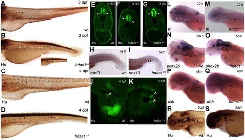
Sensory, enteric and sympathetic neuron development in hdac1b382 mutants. A–D Anti-Hu/16A11 staining of wild-type and hdac1b382 mutants at 3dpf and 4dpf. Black arrowheads indicate normally positioned DRG in wild-type and hdac1b382 mutants. White arrowheads indicate examples of DRG neurons in ectopic locations. Black asterisks (A-D and inset) indicate the regions in the tail of wild-type and hdac1b382 mutants where DRG are present. White asterisks (B and inset, D) indicate the regions in the tail of hdac1b382 mutants where DRG are absent. E-G a cross sections of wild-type (E) and examples of 2 sections (F, G) of hdac1b382 mutants illustrating that DRG neurons in mutant embryos are often ectopically localized. H, I wild-type and hdac1b382 mutants with sox10 expression in the tail region at 52 hpf, black arrowheads in I indicate distinct foci of sox10 expression in mutant in locations where DRG would normally reside. J, K cross-sections of the anterior gut in wild-type and hdac1b382 mutants stained with anti-Hu/16A11 labeling enteric neurons at 4 dpf (white arrowheads). L, M th expression in the cervical sympathetic ganglion (black arrowhead) in wild-type and hdac1b382 mutants at 72 hpf. N, O, phox2b labeling of sympathetic neuron precursors (black arrowheads) in wild-type and hdac1b382 mutants at 56 hpf. P, Q, dbh staining of sympathetic neurons (black arrowheads) in wild-type and hdac1b382 mutants at 48 hpf. R, S, Anti Hu/16A11 staining of wild-type and hdac1b382 mutants at 3dpf with black arrowheads indicating sympathetic neurons.
|