
Fig. 4
- ID
- ZDB-FIG-130618-32
- Publication
- Hashiguchi et al., 2013 - Anteroposterior and dorsoventral patterning are coordinated by an identical patterning clock
- Other Figures
- All Figure Page
- Back to All Figure Page
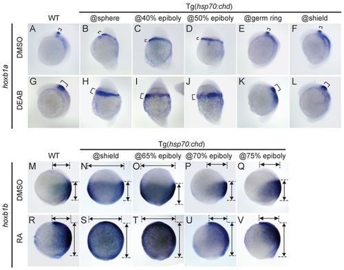
Posteriorly expanded hoxb1a and anteriorly expanded hoxb1b caused by altered RA signaling are patterned by BMP signaling with same temporal dynamics as the normal domains. (A-L) Expression of hoxb1a (bracket) in wild type (A,G) and in Tg(hsp70:chd) embryos subject to HS at the indicated stages (B-F,H-L), without (A-F) and with (G-L) DEAB treatment to inhibit RA signaling. (M-V) Expression of hoxb1b in wild type (M,R) and in Tg(hsp70:chd) embryos subject to HS at the indicated stages (N-Q,S-V), without (M-Q) and with (R-V) RA treatment. DEAB and RA treatments began at shield stage. Embryos are shown at (A-L) 6-somite and (M-V) 90% epiboly stages. Lateral views, dorsal to right. A, n=19/19; B, n=11/11; C, n=10/11; D, n=9/10; E, n=10/12; F, n=16/16; G, n=19/23; H, n=20/24; I, n=19/25; J, n=25/27; K, n=20/23; L, n=21/25; M, n=14/14; N, n=19/22; O, n=19/20; P, n=22/24; Q, n=24/24; R,=20/24; S, n=17/20; T, n=20/23; U, n=21/22; V, n=17/20. |