
Fig. 5
- ID
- ZDB-FIG-130618-33
- Publication
- Hashiguchi et al., 2013 - Anteroposterior and dorsoventral patterning are coordinated by an identical patterning clock
- Other Figures
- All Figure Page
- Back to All Figure Page
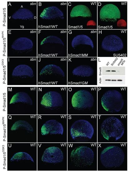
Localization of P-Smad1/5MAPK, P-Smad1/5GSK3 and total Smad1/5 during gastrulation. (A-K) Localization of P-Smad1/5 (A, n=4/4; B, n=5/6), total Smad1/5 (C, n=6/6; D, n=5/5), P-Smad1/5MAPK (E, n=5/5; F, n=4/5; G, n=3/3; H, n=7/7) and P-Smad1/5GSK3 (I, n=3/3; J, n=4/4; K, n=2/3) in sbndtc24 mutants (A,B,E-G,I-K) or wild type (C,D,H) at early gastrula stage (A-C,E-G,I-K), 75% epiboly (D) or 60% epiboly (H) stage. Embryos were injected with hSmad1WT (B,F,J), hSmad1MM (G) or hSmad1GM (K) RNA or were treated with SU5402 (H). (L) Western blot of total Smad5 protein in wild-type embryos and those treated with SU5402 or injected with smad5 MOs. Embryos were collected at 65% epiboly. Actin is a loading control. (M-X) Localization of P-Smad1/5 (M-P), P-Smad1/5MAPK (Q-T) and P-Smad1/5GSK3 (U-X) in wild type at shield (early gastrula) (M, n=8/8; Q, n=10/12; U, n=9/10), 70% epiboly (N, n=6/6; R, n=9/13; V, n=3/5), 80% epiboly (O, n=7/7; S, n=12/15; W, n=4/5) and 90% epiboly (late gastrula) (P, n=7/7; T, n=8/11; X, n=3/5) stage. All are merged confocal images of P-Smad1/5 (green), total Smad1/5 (green), P-Smad1/5MAPK (green), P-Smad1/5GSK3 (green) and DAPI (blue, except red in for C,D). Lateral views, dorsal to right. A, animal pole; Vg, vegetal pole; V, ventral; D, dorsal. |