
|
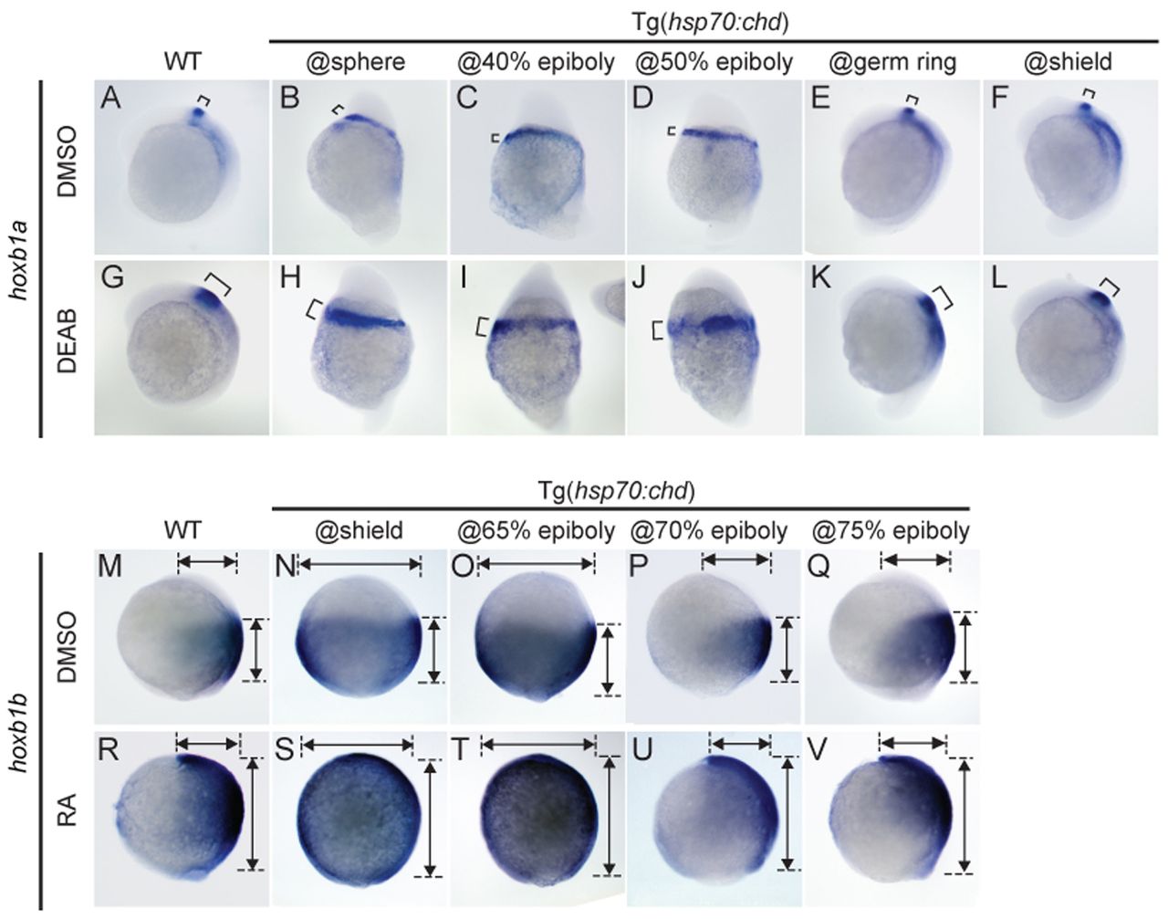
Fig. 4 Posteriorly expanded hoxb1a and anteriorly expanded hoxb1b caused by altered RA signaling are patterned by BMP signaling with same temporal dynamics as the normal domains. (A-L) Expression of hoxb1a (bracket) in wild type (A,G) and in Tg(hsp70:chd) embryos subject to HS at the indicated stages (B-F,H-L), without (A-F) and with (G-L) DEAB treatment to inhibit RA signaling. (M-V) Expression of hoxb1b in wild type (M,R) and in Tg(hsp70:chd) embryos subject to HS at the indicated stages (N-Q,S-V), without (M-Q) and with (R-V) RA treatment. DEAB and RA treatments began at shield stage. Embryos are shown at (A-L) 6-somite and (M-V) 90% epiboly stages. Lateral views, dorsal to right. A, n=19/19; B, n=11/11; C, n=10/11; D, n=9/10; E, n=10/12; F, n=16/16; G, n=19/23; H, n=20/24; I, n=19/25; J, n=25/27; K, n=20/23; L, n=21/25; M, n=14/14; N, n=19/22; O, n=19/20; P, n=22/24; Q, n=24/24; R,=20/24; S, n=17/20; T, n=20/23; U, n=21/22; V, n=17/20.