
Fig. 4
- ID
- ZDB-FIG-130510-33
- Publication
- Nevis et al., 2013 - Tbx1 is required for second heart field proliferation in zebrafish
- Other Figures
- All Figure Page
- Back to All Figure Page
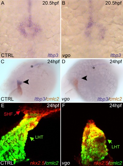
Initial specification of SHF progenitors is not perturbed in vgo embryos. A,B: ltbp3 was observed via whole-mount in situ hybridization at 20.5hpf/23ss in control (A) and vgo (B) embryos (n>12). Dorsal view, anterior down. 20× magnification. C,D: Whole-mount double in situ hybridization at 26 hpf shows ltbp3+ (blue) cells at the arterial pole (arrowhead) of the cmlc2+ (red) heart tube in control embryos. ltbp3 expression is drastically reduced (21%) or absent (79%) at the arterial pole of vgo hearts (n=14). Asterisk indicates ltbp3 expression within the notochord. E,F: At 26hpf, double transgenic Tg(nkx2.5::ZsYellow); Tg(cmlc2::GFP) control (E) and vgo (F) embryos were co-immunostained with GFP antibody (anti-GFP, green) and ZsYellow antibody (anti-RCFP, red). The future atrial segment of the linear heart tube (LHT, green arrow) expresses cmlc2 alone (green), while the future proximal ventricular myocardium co-expresses cmlc2 and nkx2.5 (yellow). Non-myocardial nkx2.5+ second heart field (SHF) progenitors (red arrow) can be visualized in control animals (n=8), but are lacking in vgo mutants (n=7). |
| Genes: | |
|---|---|
| Fish: | |
| Anatomical Terms: | |
| Stage Range: | 20-25 somites to Prim-5 |