
Fig. 1
- ID
- ZDB-FIG-130510-30
- Publication
- Nevis et al., 2013 - Tbx1 is required for second heart field proliferation in zebrafish
- Other Figures
- All Figure Page
- Back to All Figure Page
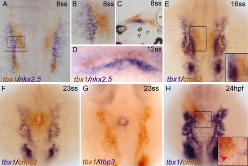
tbx1 transcripts do not co-localize with CPCs or cardiomyocytes during early cardiogenesis in zebrafish. Whole-mount double in situ hybridizations are shown. A–D: tbx1 (red) and nkx2.5 (blue) transcripts are non-overlapping at 8ss and 12ss. A: Flat mount, dorsal view, anterior up, 10× maginification. B: 20× magnification of the ALPM (boxed region in A). C: Transverse cryo-section, through the left ALPM (location of section shown by the solid line in A), 20× magnification, nt = neural tube, nc = notochord, Y = yolk. D: Dorso-lateral view showing that tbx1-expressing cells reside dorsal to nkx2.5-expressing CPCs at 12ss. E,F: tbx1+ cells are lateral to cmlc2+ cardiomyocytes at 16ss and 23ss. Dorsal view, anterior up, 10× magnification. Inset is 20× magnification of the boxed region photographed from a more dorsolateral view. G: tbx1+ cells are lateral to ltbp3+ SHF progenitors at 20.5hpf/23ss. Dorsal view, anterior up, 10× magnification. H: At 24hpf, the linear heart tube resides dorsal to the tbx1 expression domain in different planes of focus. Dorsal view, anterior up, 10× magnification. tbx1 is not expressed at the arterial pole of the linear heart tube where SHF progenitors reside (red arrowhead). Inset is 20× magnification of boxed region. |
| Genes: | |
|---|---|
| Fish: | |
| Anatomical Terms: | |
| Stage Range: | 5-9 somites to Prim-5 |