
Fig. 4
- ID
- ZDB-FIG-130503-16
- Publication
- Amali et al., 2013 - Zebrafish hoxd4a Acts Upstream of meis1.1 to Direct Vasculogenesis, Angiogenesis and Hematopoiesis
- Other Figures
- All Figure Page
- Back to All Figure Page
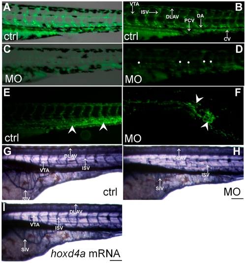
Loss of hoxd4a function impairs development of the vasculature. (A–D) Fluorescent images of the trunk and tail regions of Tg(fli1:EGFP) embryos at 48 hpf. The panels present merged bright field and fluorescent images (A,C) or fluorescent images only (B,D) The normal pattern of the vasculature (A,B) is severely disrupted in hoxd4a morphants (C,D) Dorsal extremities of ISV sprouts that fail to contact the DLAV are marked by white dots (D). The caudal vein plexus of control embryos (E, arrowheads) is replaced by a disorganized mass of endothelial tissue in hoxd4a morphants (F, arrowheads). (G–I) Alkaline phosphatase staining at 72 hpf revealing the vasculature in (G) control-injected larvae, (H) hoxd4a morphants, and (I) rescued larvae co-injected with capped mRNA for hoxd4a. Dorsal aorta (DA), posterior cardinal vein (PCV), inter-segmental vessels (ISV), caudal artery (CA), dorsal longitudinal anastomotic vessel (DLAV), caudal vein (CV) and vertebral artery (VTA). All images show lateral views, with anterior to the left and dorsal on top. Scale bars equal 100 μm. |
| Fish: | |
|---|---|
| Knockdown Reagent: | |
| Observed In: | |
| Stage Range: | Long-pec to Protruding-mouth |