
FIGURE
Fig. 1
- ID
- ZDB-FIG-130502-3
- Publication
- Muto et al., 2013 - Real-Time Visualization of Neuronal Activity during Perception
- Other Figures
- All Figure Page
- Back to All Figure Page
Fig. 1
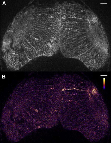
Spontaneous Neuronal Activity in the Optic Tectum in a Zebrafish Larva(A) UAS:GCaMP7a fish were mated to gSA2AzGFF49A fish that expressed Gal4FF in the tectum. A double-transgenic larva at 3 dpf was embedded in agarose and imaged with a confocal microscope. A single raw image of the fluorescence intensity extracted from a time-lapse movie is shown (see Movie S1).(B) The ratiometric image of (A) was created and pseudocolored to reveal the fluorescence change in the cell body and axon neurite of a single neuron.Scale bars represent 20 μm. |
Expression Data
| Gene: | |
|---|---|
| Fish: | |
| Anatomical Terms: | |
| Stage: | Protruding-mouth |
Expression Detail
Antibody Labeling
Phenotype Data
Phenotype Detail
Acknowledgments
This image is the copyrighted work of the attributed author or publisher, and
ZFIN has permission only to display this image to its users.
Additional permissions should be obtained from the applicable author or publisher of the image.
Full text @ Curr. Biol.