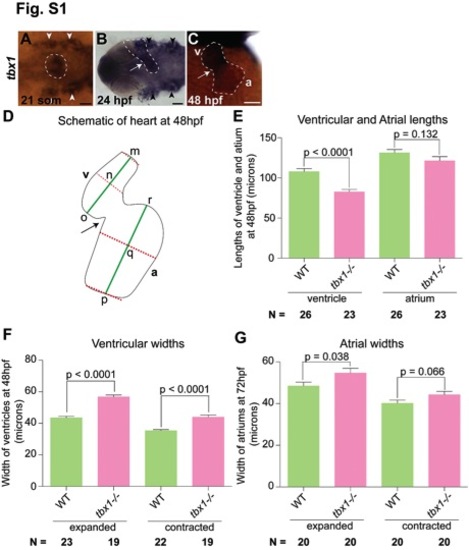
tbx1 expression and heart shape defect of tbx1-/- 1arvae. ISH for tbx1 in WT embryos showing expression in the fusing heart fields (21 somites, A), linear heart tube (24 hpf, B) and the looped heart (48 hpf, C). A and B are dorsal views and C is a head-on view. Arrowheads in A and B point towards pharyngeal pouches and the cardiac cells are outlined in A–C. (D) Schematic of head-on view of 48 hpf heart. Arrow indicates the AVC; a, atrium; v, ventricle. Dotted red lines indicate the widths at the ends and at the widest part of the ventricle and atrium. The longitudinal axes (shown in green) were drawn by joining the midpoints m and n in ventricle and midpoints p and q in atrium. mo and pr were measured for length of ventricle and atrium, respectively (see Materials for details). (E) Measurement of lengths of the ventricle and atrium in WT and tbx1-/- mutants. (F, G) Measurement of the widths of the ventricles at 48 hpf and atriums at 72 hpf in tbx1-/- mutants and WT siblings in the expanded and contracted states. WT siblings are represented in green and tbx1-/- mutants in pink. N indicates the total number of embryos represented in the plot. Scale bars: 50 μm.
|
