|
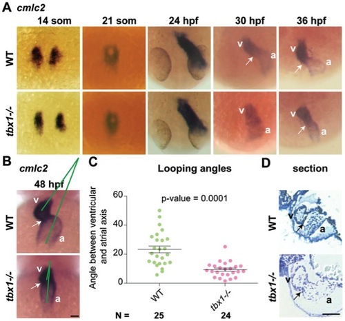
tbx1 expression and heart looping defects in tbx1-/- embryos. (A) cmlc2 ISH in tbx1-/- mutants and WT siblings at specification (14 somites), fusion (21 somites), linear heart tube (24 hpf), jogging (30–36 hpf) stages. (B) By 48 hpf the heart has finished looping in WT forming an acute angle between the atrial and ventricular axes (green lines), while in tbx1-/- mutants the axes are nearly parallel. (C) Comparison of atrio-ventricular axis angles between wild-type (left, green) and tbx1-/- mutants (right, red). N indicates the total number of embryos represented in the plot. (D) 5 micron sections of the hearts from 72 hpf embryos, showing wider ventricle and atrium and thinner heart walls in tbx1-/- mutant as compared to WT sibling. Arrows indicate the AVC; a, atrium; v, ventricle. Scale bar: 50 μm.
|