|
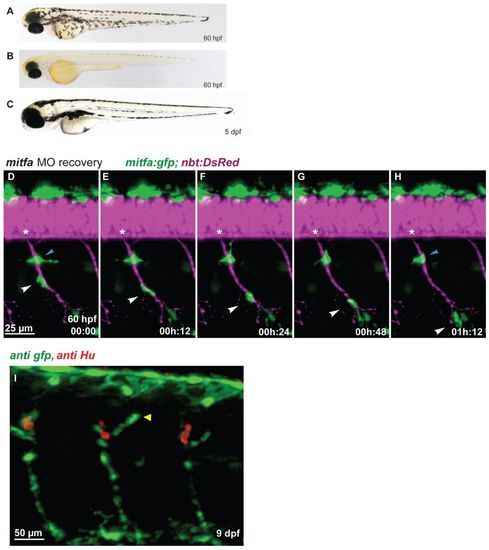
Regenerating MPs emerge at the site of the DRGs and travel along the spinal nerves. (A-C) In contrast to wild type (A), mitfa-MO-treated zebrafish embryos (B) lack larval melanophore pigmentation until 60 hpf, but regenerate the larval melanophore stripes completely by day 5 (C). (D-H) Tg(mitfa:gfp; nbt:DsRed) embryo previously treated with mitfa-MO imaged for several hours starting at 60 hpf (digital sectioning). A GFP-positive cells (white arrowhead) migrates along the spinal nerve. Another cell remained stationary (blue arrowhead) but was later observed to migrate away. Asterisks indicate the position of the DRG. (I) In mitfa morphant larvae (9 dpf), the regenerating MPs form a string of mitfa-positive cells along the spinal nerves associated with the DRGs both dorsally (arrowhead) and ventrally.
|