
Fig. 5
- ID
- ZDB-FIG-130211-6
- Publication
- Nair et al., 2013 - In vitro oocyte culture-based manipulation of zebrafish maternal genes
- Other Figures
- All Figure Page
- Back to All Figure Page
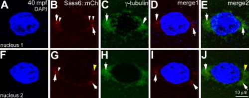
Sass6-mCherry protein from exogenous mRNA injected into stage IV oocytes localizes to centrioles during early cleavage stages. Immunolabeling for γ-tubulin and DAPI staining for nuclei in a Sass6-mCherry expressing blastodisc during prophase (40 mpf), which contains two nuclei, both of which are shown. C,E,H,J: γ-tubulin labels pericentriolar material around the nuclei, with intense foci in presumptive centrioles flanking each nuclei (arrows). B,D,E,G,I,J: Ectopically expressed Sass6-mCherry protein also localizes to foci flanking the nuclei (arrows in B,D,G,I) and colocalizes with γ-tubulin foci (arrows in E,J). B,G: Ectopic Sass6-mCherry is also observed as additional foci near the nucleus periphery (white arrowheads in B,G) and in the cytoplasm (not shown), consistent with the production of supernumerary centrioles after overexpression of maternally-derived Dsas-6 in Drosophila embryos (Peel et al., 2007). G,J: The observed putative supernumerary foci can also be associated with extra γ-tubulin foci (yellow arrowhead). Merge1: DAPI and Sass6::mCherry; merge2: DAPI, Sass6::mCherry and γ-tubulin. |