
Fig. S4
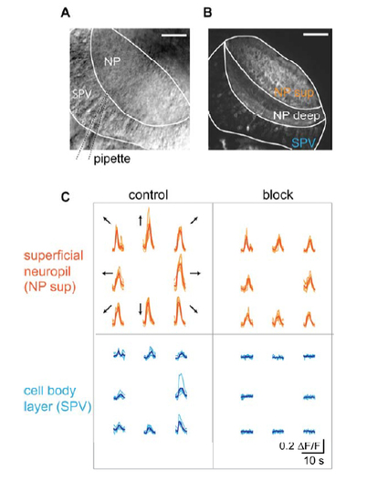
Pharmacological block of postsynaptic activity (A) Dorsal view of a tectal hemisphere imaged with an infrared-sensitive transillumination dectector below the recording chamber. Tectal neuropil (NP) and cell body layer (SPV: stratum periventriculare) are visible, as well as the broken patch pipette (dashed lines) that is used to locally apply inhibitors of ionotropic 9 glutamate receptor channels by weak positive pressure. Rostral is to the top. Scale bar: 40 µm. (B) Dorsal view of a tectal hemisphere imaged in a Tg(huC:Gal4;UAS:GCaMP3) animal showing neuropil structure (NP sup: superficial neuropil innervated by RGC axons; NP deep: deep neuropil) and cell body layer (SPV). Scale bar: 40 µm. (C) Ca2+ transients from the regions shown in (B) during presentation of bars moving in eight directions before (“control”) and during local application of 20-100 µM APV and 10-50 µM CNQX (“block”) by maintaining weak positive pressure in the application pipette for several minutes. |