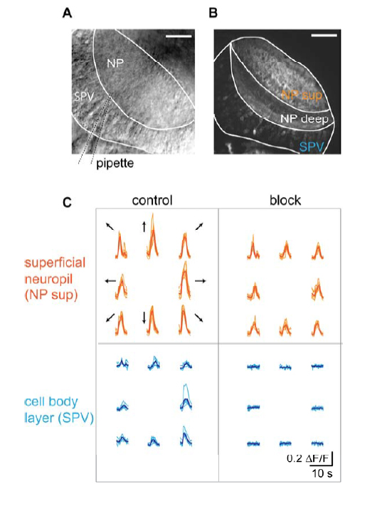
Fig. S4 Pharmacological block of postsynaptic activity (A) Dorsal view of a tectal hemisphere imaged with an infrared-sensitive transillumination dectector below the recording chamber. Tectal neuropil (NP) and cell body layer (SPV: stratum periventriculare) are visible, as well as the broken patch pipette (dashed lines) that is used to locally apply inhibitors of ionotropic 9 glutamate receptor channels by weak positive pressure. Rostral is to the top. Scale bar: 40 µm. (B) Dorsal view of a tectal hemisphere imaged in a Tg(huC:Gal4;UAS:GCaMP3) animal showing neuropil structure (NP sup: superficial neuropil innervated by RGC axons; NP deep: deep neuropil) and cell body layer (SPV). Scale bar: 40 µm. (C) Ca2+ transients from the regions shown in (B) during presentation of bars moving in eight directions before (“control”) and during local application of 20-100 µM APV and 10-50 µM CNQX (“block”) by maintaining weak positive pressure in the application pipette for several minutes.
Image
Figure Caption
Acknowledgments
This image is the copyrighted work of the attributed author or publisher, and
ZFIN has permission only to display this image to its users.
Additional permissions should be obtained from the applicable author or publisher of the image.
Full text @ Neuron

