
Fig. S1
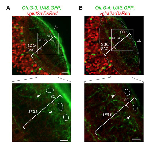
Characterization of novel Gal4 lines (A) Tg(Oh:G-3;UAS:GFP) fish were crossed with Tg(vglut2a:DsRed) fish to obtain triple transgenic animals (optical section from a confocal stack; dorsal view). In the SO, superficial interneurons (SINs) can be detected as dark shadows, serving as landmarks (dashed outlines) (Del Bene et al., 2010). GFP-positive processes are present near the SFGS/SO border (filled arrowheads in lower panel). In this line, 3 there is bright skin fluorescence (open arrowhead, upper panel). Scale bar: 20 µm (upper panel) / 10 µm (lower panel). (B) In Tg(Oh:G-4;UAS:GFP;vglut2a:DsRed) animals, GFP-labeled processes are absent from the SFGS/SO border (filled arrowheads in lower panel). Scale bar: 20 µm (upper panel) / 10 µm (lower panel). Skin fluorescence marked by open arrowhead (upper panel). |