
Fig. 5
- ID
- ZDB-FIG-130118-14
- Publication
- Freitas et al., 2012 - Hoxd13 contribution to the evolution of vertebrate appendages
- Other Figures
- All Figure Page
- Back to All Figure Page
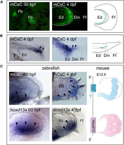
Enhancer Activity of Mouse CsC in Zebrafish Fins Developmental stages are on upper left corners in each panel.(A) Enhancer activity of mouse CsC (mCsC) shown by the expression of GFP in the posterior half of the fin buds (Fb) at 30 hpf and surrounding the distal margin of the endoskeletal disc 4 dpf. Bracket line delimits the distal border of this territory. The scheme on the right represents the main domain of mCsC activity in zebrafish fins at 4 dpf (green).(B) Transverse sections throughout a mCsC transgenic fin expressing GFP at 4 dpf. Left panel shows restriction of GFP mRNA to the distal tip of the endoskeletal territory (arrowheads). Central panel shows a higher magnification of the expressing region (arrowheads). Note GFP expression in chondrocyte-like cells (delimited by a white dashed line) and in surrounding cells. The scheme on the right represents the inferred localization of the expression pattern in the endoskeletal territory (green: chondrocytes and undifferentiated distal mesenchyme).(C) Comparison between mCsC activity and hoxd13 expression in zebrafish and mouse. Dashed lines indicate the distal limits of the endoskeletal territory. Left panel shows that expression of GFP in mCsC transgenic zebrafish fin is nested within hoxd13a expressing domain at 60 hpf (arrowheads). Central panel shows simultaneous anterior expansion of mCsC activity and hoxd13a expression throughout the distal portion of the endoskeletal territory at 4 dpf (arrowheads). Note that mCsC activity is detected also within a subdomain of hoxd13a expression at this stage. Schemes on the right are based on Gonzalez et al. (2007). Left scheme indicates relative position of mCsC upstream of Hoxd13 in mice. Schemes on the right represent CsC and Hoxd13 expression in mouse limbs (blue and pink respectively). Note that mCsC is also active in a subdomain of Hoxd13 expression in this organism. |
Reprinted from Developmental Cell, 23(6), Freitas, R., Gómez-Marín, C., Wilson, J.M., Casares, F., and Gómez-Skarmeta, J.L., Hoxd13 contribution to the evolution of vertebrate appendages, 1219-1229, Copyright (2012) with permission from Elsevier. Full text @ Dev. Cell