
Fig. 3
- ID
- ZDB-FIG-130118-12
- Publication
- Freitas et al., 2012 - Hoxd13 contribution to the evolution of vertebrate appendages
- Other Figures
- All Figure Page
- Back to All Figure Page
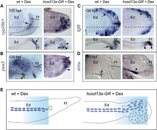
Other Distal Markers Are Also Affected by hoxd13a Overexpression Each panel shows expression pattern of a gene at 120 hpf, in which wild-type controls (WT + Dex) and Dex-treated hoxd13a-GR injected fins are on left and right columns, respectively. Stages of development are indicated at left of each panel. Arrows point to the WT expression and arrowheads indicate expression induced by hoxd13a-GR injections. Ed, endoskeletal disc; Ff, finfold.(A and B) cyp26b1 (A) and pea3 (B) is extended distally in hoxd13a overexpressing fins. Dashed lines in (A) indicate the approximate plane of the sections shown bellow. In these sections distal cyp26b1 expression can be observed surrounding the differentiated chondrocytes (yellow dashed line) in injected fins.(C) fgf8a expression is confined to a narrower distal domain in injected fins. Dashed lines indicate the approximate plane of the section shown bellow.(D) shha is upregulated in hoxd13a overexpressing fins.(E) Schematic representations of the observed phenotypes. Blocks of graded blue represent the domain where characteristic distal limb markers (hox13b, pea3, and cyp26b1) are expressed in wild-type and in hoxd13a overexpressing fins. |
Reprinted from Developmental Cell, 23(6), Freitas, R., Gómez-Marín, C., Wilson, J.M., Casares, F., and Gómez-Skarmeta, J.L., Hoxd13 contribution to the evolution of vertebrate appendages, 1219-1229, Copyright (2012) with permission from Elsevier. Full text @ Dev. Cell