
Fig. 2
- ID
- ZDB-FIG-121212-36
- Publication
- Gyda et al., 2012 - The tumor suppressor gene retinoblastoma-1 is required for retinotectal development and visual function in zebrafish
- Other Figures
- All Figure Page
- Back to All Figure Page
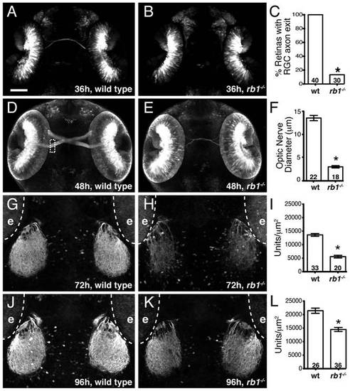
Rb1-deficient embryos possess delayed retinotectal projection. Dorsal view of maximum intensity projection of confocal z-stacks of RGC axons in wild type or rb1te226; ath5:gfp+/- embryos. (A–C) RGC axons fail to exit rb1te226 retina at 36 hpf. (D–F) rb1te226 optic nerves are hypoplastic. Diameter measured upon retinal exit, marked by dashed rectangle. (G-L) Tectal innervation is reduced in rb1te226 at 72 (G-I) and 96 hpf (J–L). Innervation measured as a function of pixel intensity units per µm2 from summation intensity projections of tectal area (not shown, see Experimental Procedures). Digitally excluded eyes (e) are outlined by dotted line. (C, F, I, L) Bar graphs represent the mean with error bars denoting SEM. *p<0.001; one-way ANOVA (F, I, L) or binomial z-test (C). N retinas, optic nerves, or tecta shown at base of bar graphs. Anterior to the top of panel, scale bar = 50 μm. |
| Fish: | |
|---|---|
| Observed In: | |
| Stage Range: | Prim-25 to Day 4 |