
Fig. 9
- ID
- ZDB-FIG-121031-41
- Publication
- Shimizu et al., 2012 - Visualization and exploration of Tcf/Lef function using a highly responsive Wnt/beta-catenin signaling-reporter transgenic zebrafish
- Other Figures
- All Figure Page
- Back to All Figure Page
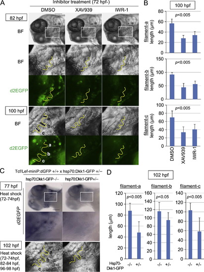
Wnt/β-catenin signaling was required for gill filament growth. (A–D) XAV939 or IWR-1 treatment or induction of Dkk1-GFP expression blocked Tcf/Lef-miniP:dGFP reporter activity in gill filament buds and reduced the gill filament length. (A, B) Line-2 embryos were treated with DMSO, 10 μM XAV939, or 10 μM IWR-1 from 72 hpf. Panels show lower left-side head views (top panels) and magnified gill area views (second, third, forth, and bottom panels) of 82- and 100-hpf inhibitor-treated Line-2 embryos, with the anterior side to the left. Insets in the top panels indicate the gill areas shown in the second and third panels. Bright-field (BF) images are shown in the top, second, and fourth panels in (A). d2EGFP expression was observed by fluorescence microscopy (third and bottom panels in A). The outlines of the gill filaments and their buds are shown with yellow broken lines. Scale bar, 100 μm. (C, D) hsp70:Dkk1-GFP-carrying Line-2 embryos were generated and classified using methods similar to those shown in Fig. 2(A). hsp70:Dkk1-GFP carriers (+/-) or non-carriers (-/-) were exposed to heat shock for 2 h at 37 °C at 72 hp. d2EGFP expression was detected by in situ hybridization 3 h after heat shock (top and middle panels in C). Panels show lower left-side head views (top panels) and magnified gill area views (middle and bottom panels) for 77- and 102-hpf inhibitor-treated Line-2 embryos, with the anterior side to the left. Insets in the top panels indicate the gill areas shown in the second panels. The embryos shown in the bottom panels were exposed to two additional heat shock treatments for 2 h at 37 °C at 82 and 96 hpf. The outlines of the gill filaments and their buds are shown with yellow broken lines. Scale bar, 100 μm. Three gill filaments that budded from branchial arch 1 are labeled as a, b, and c, and their lengths are shown. (B, D) Graphs showing the lengths of the gill filaments indicated in (A) and (C) for 100-hpf inhibitor-treated reporter fish embryos (B, n=10 each) or 102-hpf heat shock-exposed embryos (D, n=11 each). Data are mean±SD. The p value was calculated by Student′s t test. The gill filament length in 100-hpf DMSO-treated embryos (B) was shorter than that in 102-hpf hsp70:Dkk1-GFP non-carriers (-/-). This may be due to differences in the treatment. |
Reprinted from Developmental Biology, 370(1), Shimizu, N., Kawakami, K., and Ishitani, T., Visualization and exploration of Tcf/Lef function using a highly responsive Wnt/beta-catenin signaling-reporter transgenic zebrafish, 71-85, Copyright (2012) with permission from Elsevier. Full text @ Dev. Biol.