
Fig. 8
- ID
- ZDB-FIG-121031-40
- Publication
- Shimizu et al., 2012 - Visualization and exploration of Tcf/Lef function using a highly responsive Wnt/beta-catenin signaling-reporter transgenic zebrafish
- Other Figures
- All Figure Page
- Back to All Figure Page
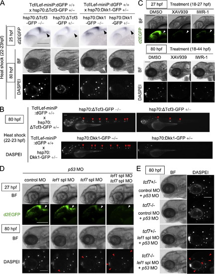
Tcf7/Lef1-mediated Wnt/β-catenin signaling was required for aLL development. (A, B) Induction of ΔTcf3-GFP or Dkk1-GFP expression blocked Tcf/Lef-miniP:dGFP reporter activity in prim-A and reduced the neuromast number. hsp70:ΔTcf3-GFP- or hsp70:Dkk1-GFP-carrying Line-2 embryos were generated and classified using methods similar to those shown in Fig. 2(A). hsp70:Dkk1-GFP carriers (+/-), hsp70:ΔTcf3-GFP carriers (+/) or non-carriers (-/-) were exposed to heat shock for 1 h at 37 °C at 22 hpf. d2EGFP expression was detected by in situ hybridization 2 h after heat shock (top panels in A). Neuromasts were visualized at 80 hpf by DASPEI staining (A, B). Panels show head (A) or entire (B) views of embryos, with the anterior side to the left. Bright-field (BF) images are shown in the middle panels (A). DASPEI-stained cells were observed by fluorescence microscopy (bottom panels in A, panels in B). prim-As (A) and DASPEI-stained prim-I-derived primary neuromasts (B) are indicated with black and red arrowheads, respectively. Scale bar, 100 μm. (C) XAV939 or IWR-1 treatment reduced Tcf/Lef-miniP:dGFP reporter activity in prim-A and the number of prim-A-derived neuromasts. Panels show left-side head views of 27- and 80-hpf zebrafish embryos treated with DMSO, 10 μM XAV939, or IWR-1 from 18 to 27 hpf or 18 to 44 hpf, with the anterior side to the left. Bright-field (BF) images are shown in the top and third panels. d2EGFP-expressing cells (second panels) and DASPEI-stained cells were observed by fluorescence microscopy (bottom panels). Scale bar, 100 μm. (D, E) Tcf7 and Lef1 contributed to aLL development. Left-side views of 27- or 80-hpf Line-2 (D) heterozygous (+/-) or homozygous (-/-) tcf7 mutant (E) embryos injected with control MO, tcf7 MO, lef1 spl MO, and p53 MO, as indicated, with the anterior side to the left. Heterozygous (+/-) tcf7 mutant embryos were obtained by crossing homozygous (-/-) tcf7 mutants with wild-type zebrafish. Bright-field (BF) images are shown in the top and third panels (D) or the left panels (E). d2EGFP-expressing cells (second panels in D) and DASPEI staining (bottom panel in D and right panels in E) were visualized by fluorescence microscopy. prim-As are indicated with white arrowheads. The missing neuromasts in the morphants are indicated with red arrowheads. Scale bar, 100 μm. |
Reprinted from Developmental Biology, 370(1), Shimizu, N., Kawakami, K., and Ishitani, T., Visualization and exploration of Tcf/Lef function using a highly responsive Wnt/beta-catenin signaling-reporter transgenic zebrafish, 71-85, Copyright (2012) with permission from Elsevier. Full text @ Dev. Biol.