
Fig. S1
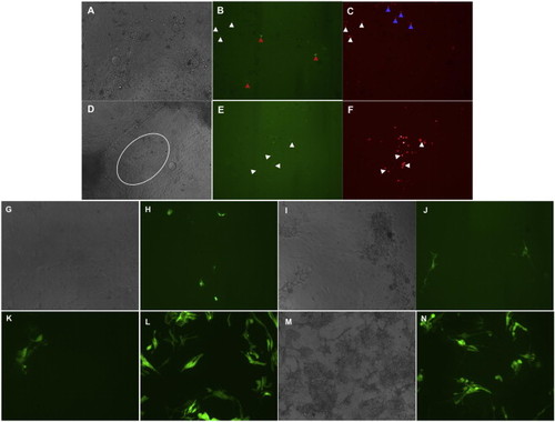
Differentiation of Primary Cells Derived from Additional Transgenic Embryos, Related to Figure 2 (A–F) Cells derived from double transgenic embryos of scl-GFP and gata1-dsRed express both markers at day 2 (A–C) and at day 9 (D–F). Scl-GFP and gata1-dsRed are colocalized in some round-shaped cells (white arrowheads in B and C), and are also exclusively expressed in some cells (GFP, non-round cells, red arrowheads in B; and DsRed, round cells, blue arrowheads in C). Gata1-DsRed-expressing round cells can survive until day 9 and form colony-like structures (white circle in D and red round cells in F) with stronger expression, some of which overlie Scl-GFP-expressing cells (white arrowheads in E and F). (G–N) Cmlc2-GFP transgene marks myocardium at day 2 (G and H) and day 5 (I and J), with some contracting cells. The transgene MLC-GFP shows expression in elongated and flat muscle cells at day 2 (K) and day 3 (L), when culture begins at the dome stage, and at day 4 (M, bright field, and N), starting to culture from shield-stage embryo cells. |