
Fig. 6
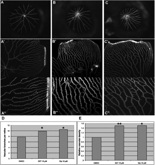
Test of Selected Candidate Compounds in Vivo (A–C) Representative images of retinal vasculature treated with DMSO, SST, and Dip. (A, A2, and A3) 0.5% DMSO. (B, B2, and B3) 10 μM SST. (C, C2, and C3) 10 μM Dip. (A–C) Frontal view of retinal vasculature after removal of cornea and lens of Flk1-GFP transgenic adult eyes without flat mount. (A2–C3) Frontal view after flat mount. (A3, B3, and C3) Higher magnification of A2, B2, and C2, respectively. (D and E) Quantifications of vessel number and relative density per eye, respectively. p = 0.0216 for SST and p = 0.022 for Dip against control in vessel numbers. p = 0.009 for SST and p = 0.025 for Dip against control in relative density. |