
Fig. 2
- ID
- ZDB-FIG-121016-2
- Publication
- McGraw et al., 2012 - Postembryonic neuronal addition in Zebrafish dorsal root ganglia is regulated by Notch signaling
- Other Figures
- All Figure Page
- Back to All Figure Page
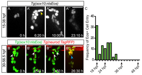
Neural crest migration into the dorsal root ganglia (DRG) ceases prior to neuron differentiation. (A-A′′ ′) Confocal projections of stills from a time-lapse movie showing migrating Tg(sox10:nlsEos) + neural crest cells between 15 and 38 dpf. (A) At time zero, the region of DRG formation contains two Eos + cells. (A′) A still at 6:20 hours shows a representative neural crest cell joining the DRG anlagen (yellow arrowhead). Between 10:00 hours (A′′) and 23:10 hours (A′′ ′), no neural crest cells migrate into the DRG. (B-B′′ ′) Time-lapse movie a Tg(sox10:nlsEos)/Tg(neurod:TagRFP) transgenic embryo between 30 and 57:10 hpf. (B) An Eos+/RFP- cell (white arrowhead) at time zero, begins to express RFP by 0:40 hours (B′) and divides at 0:50 hours (B′′). (B′′ ′) After 26.30 hours of imaging, neurons are labeled with RFP (blue arrow). (C) Quantification of Eos + cells that migrate into the DRG anlagen between 18 and 48 hpf. No new neural crest cells join the DRG after 32 hpf (n = five embryos). Scale bars, 25 μm. dpf, days postfertilization; hpf, hours postfertilization. |