
Fig. 1
- ID
- ZDB-FIG-121016-1
- Publication
- McGraw et al., 2012 - Postembryonic neuronal addition in Zebrafish dorsal root ganglia is regulated by Notch signaling
- Other Figures
- All Figure Page
- Back to All Figure Page
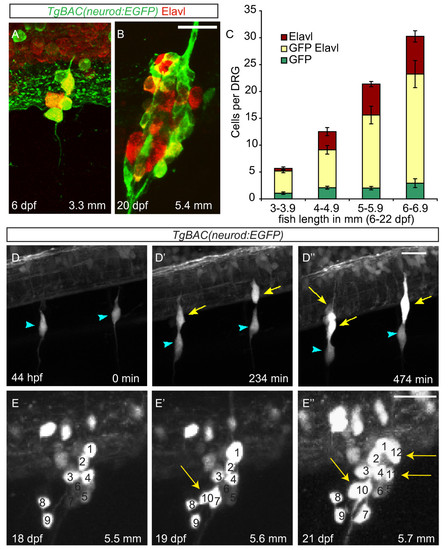
Neurons are continuously added to the larval dorsal root ganglia (DRG). (A-B) Confocal images of a TgBAC(neurod:EGFP) transgenic larva between six (A) and twenty-two dpf (B) showing DRG growth. Neurons were labeled with anti-Elavl (red) and GFP (green) antibodies. (C) Quantification (±SEM) of Elavl and GFP-positive cells per DRG in larvae staged by overall body length in millimeters (mm). Elavl-positive neurons are continuously added to the DRG as the larvae develop. Over half of the Elavl-labeled cells are also labeled with GFP (n = between five and fifteen larvae per condition, four ganglia per larva). (-D′′) Stills from a time-lapse movie showing neuron differentiation in a TgBAC(neurod:EGFP) embryo between 44 and 52 hpf. (D) At 44 hpf the DRG contain a single neuron each (blue arrowheads). (D′) By 234 minutes, neurons have been added to each ganglion (yellow arrows). (D′′) At 474 minutes the ganglia contain several neurons. (E-E′′) Analysis of a single ganglion in a TgBAC(neurod:EGFP) transgenic larval fish between 18 and 21 dpf. (E) At eighteen dpf, the ganglion contains nine GFP-positive cells. (E′) At 19 dpf, the ganglion contains 10 GFP-positive cells (white arrow) and by 21 dpf (E′′) 12 GFP-positive cells (white arrows). During the course of imaging the larval fish grew from 5.5 mm to 5.7 mm in length. Scale bars, 20 μm. dpf, days postfertilization; hpf, hours postfertilization; GFP, green fluorescent protein. |