|
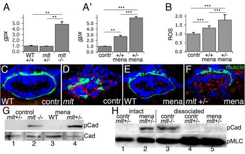
Heterozygous mlt larvae are sensitized to oxidative stress. (A) Quantitative RT-PCR shows increased intestinal gpx expression in 74 hpf mlt homozygotes (mlt -/-) compared with WT (+/+) and mlt heterozygotes (mlt +/-) (** p<.01). (A′) Quantitative RT-PCR shows intestinal gpx expression is increased in menadione treated homozygous WT larvae (+/+ mena) compared with untreated WT (contr). A much stronger response to menadione is seen in mlt heterozygotes (+/- mena) (*** p<.001). (B) ROS production in intestinal epithelial cells of 76 hpf WT control larvae (contr) versus menadione treated homozygous WT and heterozygous mlt larvae (*** p<.001). Bar graphs in (A) and (A′) show mean and standard deviation of three independent experiments. Bar graph in (B) shows mean of six larvae for each genotype; 15–25 cells per larva. (C–F) Histological cross-sections of larvae processed for fluorescent RNA in situ hybridization. Menadione induced gpx expression (red) in the epithelium but not smooth muscle (green) of heterozygous mlt larvae (F). This is comparable to the gpx expression pattern in control homozygous mlt larvae (D). (G) Western blot showing premature h-CaD phosphorylation (pCad) in dissected intestines from menadione treated heterozygotes but not homozygous WT larvae (lane 4 versus lane 3; ratio phospho-h-CaD WT:mlt = 0.02/1, relative to total h-CaD; CaD; in six experiments the ratio averaged 0.016/1; p<.001). h-CaD is prematurely phosphorylated in intestines dissected from 74 hpf mlt homozygotes versus mlt heterozygotes (lane 1 versus lane 2). (H) Western blot showing h-CaD phosphorylation (pCad) in the menadione treated heterozygous intestines prior to dissociation (lane 2) but not after dissociation into free cell populations (lane 5). Phospho-h-CaD persists in dissociated cells from homozygous intestines (lane 3), but is not detected in control intestines dissected from mlt heterozygotes, before (lane 1) or after (lane 4) dissociation into free cell populations. No phopho-h-CaD was detected in any dissociated samples in three independent experiments. Loading control, phospho-Myosin light chain (pMlc).
|