|
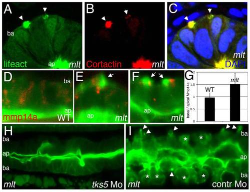
Actin rich protrusions in mlt are invadopodia homologs. (A–C) Histological cross-sections through the posterior intestine of 74 hpf mlt mutant larvae immunostained with antibodies against GFP (green) (labeling Lifeact-GFP) and Cortactin (red). DAPI-stained nuclei, blue. Lifeact-GFP and Cortactin co-localize in actin rich basal membrane protrusions (arrowheads) and in the apical brush border (ap). (D–G) Sagittal confocal scans through the intestine of 74 hpf Lifeact-GFP transgenic wild type (WT) and mlt larvae. Lifeact-GFP binds actin in the apical brush border (ap) of WT (D) and mlt (E, F) epithelial cells, as well as basal (ba) invadopodia-like protrusions in mlt (E, F, arrows). In WT, the Mmp14a-mCherry fusion protein (red) is distributed throughout the epithelial cells. In mlt, Mmp14a-mCherry is preferentially localized to the basal region of the epithelial cells. (n = 33 WT and 33 mlt cells examined; 6 larvae each genotype). (G) Ratio of basal to apical Mmp14a-mCherry in WT versus mlt epithelial cells (error bars, standard deviation. * p<.001). (H–I) Sagittal confocal scans through the intestines of 84 hpf Lifeact-GFP transgenic mlt larvae. Invadopodia (arrowheads I) and invasive cells (asterisks I) are present in the mlt larvae injected with a control morpholino (I), but are not detected in the larvae injected with the tks5 morpholino (H).
|