
Fig. 7
- ID
- ZDB-FIG-120831-15
- Publication
- Eom et al., 2012 - Melanophore Migration and Survival during Zebrafish Adult Pigment Stripe Development Require the Immunoglobulin Superfamily Adhesion Molecule Igsf11
- Other Figures
- All Figure Page
- Back to All Figure Page
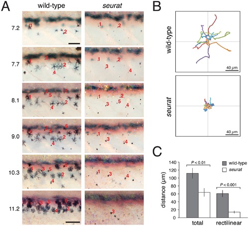
igsf11-dependent migration and survival of melanophores. (A) Repeated images of developing wild-type and seurat mutant larvae between 14–28 days post-fertilization. Numbers to the left of images are SSL. In wild-type larvae, new adult melanophores differentiated already within stripes or translocated short distances as stripes formed (e.g., note changes in the relative positions of cells 2 vs. 4, and cell 3 vs. 1 and 5). In seurat mutants, however, little movement was observed and many melanophores died as evidenced by the presence of melanized cellular debris apparent at high magnification (not shown; [32]. Images shown were rescaled to maintain the same field of view as the fish grew; scale bars at 7.2 SSL and 11.2 SSL represent 100 and 200 μm, respectively. (B) When cultured in vitro, wild-type melanophores migrated further than seurat mutant melanophores. Shown are tracks of 16 cells of each genotype. (C) Quantification of total and rectilinear distances moved by cells in vitro confirmed reduced motility of seurat mutant melanophores (t = 3.0, t = 5.4, respectively; d.f. = 26). Shown are means ± SE. |
| Fish: | |
|---|---|
| Observed In: | |
| Stage Range: | Days 14-20 to Days 21-29 |